Trending

Gastroparesis

Treatment,Gastroparesis,Gastroparesis treatment

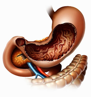
Gastroparesis is a disease that affects the muscles, or the nerves controlling muscles of the stomach. It results in improper grinding of food and delayed emptying of the stomach. In a normal person, the vagus nerve controls the muscles of the stomach which break down the food and move it further into the intestines by contracting. Any damage to the vagus nerve whether by injury or illness, will result in disruption of this function. The most common disease considered responsible for gastroparesis by nerve damage is diabetes mellitus. The second most common cause is idiopathic; no such reason can be found and it is termed as idiopathic gastroparesis. An example of the disease causing gastroparesis due to damage to stomach muscles is Scleroderma. In some cases, the problem is neither in the muscles not in the nerves; it is simply due to nerve reflex e.g. when there is pancreatitis. Apart from these, an imbalance in the levels of minerals (e.g. calcium, potassium etc) in the blood, and thyroid disease can also be responsible for causing gastroparesis.
Gastroparesis primarily presents with nausea and vomiting which mainly occurs after meals but can also be due to gastric secretions in some severe cases. These symptoms are followed by bloating, abdominal pain, abdominal distention, early satiety (patient feels full on eating small amounts of food), and weight loss. Improper or reduced food intake further leads to nutritional deficiencies.
A diagnosis, as in most diseases, is established through a thorough medical history and physical examinations, blood tests. In this case, there is a need to rule out structural problems or blockage in the GI tract. This is done through various tests including upper gastrointestinal endoscopy, upper GI series, ultrasound, gastric emptying scintipraphy, smart pill (a small electronic device that is swallowed and then moves around the GI tract and records information about the time taken for food to pass through the tract), breath test (radioactive material is ingested and breath samples are taken at regular intervals to calculate the emptying rate of the stomach). A new study known as antro-duodenal motility study has emerged that can measure the pressure generated by the contractions of the stomach and intestinal muscles. It is however still experimental and reserved for selected case. Another experimental study called electrogastrogram that works on same principle as EKG and records the signals that travel through the muscles of the stomach.
Gastroparesis treatment mainly includes diet, medication and procedures to help emptying of the stomach. The diet plan is based on foods that are easily emptied from the stomach; soft, liquid diet. The food should also be low in fiber and should mostly be eaten early during the day. The medications are given to treat the underlying conditions such as diabetes mellitus.
Medication to relieve nausea, vomiting and abdominal pain include promotility drugs (metoclopromide, domperidone, etc), anti-nausea drugs (prochlorperazine, promethazine, etc), medical marijuana (marinol), serotonin antagonists (ondansetron), and anticholinergics (scopolamine).
Drugs for reliving abdominal pain include NSAIDs (ibuprofen, naproxen, etc), tricyclic antidepressants (amitriptyline), nerve blockers (gabapentin), narcotics (tramadol, fentanyl, etc). Some drugs such as cisapride, domperidone, metoclopromide, and erythromycin are used to stimulate the contractions in the muscles of the stomach. As the stomach emptying is affected, liquid medications are preferred over oral as they are more effective in this condition.
A new method called electrical pacing is used to treat severe cases of gastroparesis. A pacemaker for the stomach is placed laproscopically and then generates impulses. Surgery can also be performed to facilitate emptying by creating a larger opening between the stomach and intestines. Sometimes, the entire stomach may be removed. However, surgery should be the last option of treatment.
Patients of gastroparesis suffer from malnutrition and dehydration which can be managed by IV fluids and replenishing of electrolytes. This involves methods such as IV total parenteral nutrition (TPN) and enteral nutrition. Enteral nutrition is preferred over TPN and can be done via naso-jejunal tubes or jejunostomy tubes. Treatment with botulinum toxin may be effective but is currently under study.

Clinical research is being conducted at National Institute of Diabetes and Digestive and Kidney Disease (NIDDK), for new medications or surgical methods that can cure gastroparesis more effectively and safely. More information can be found at cancercarevilla.blogspot.com.
Treatment,Gastroparesis,Gastroparesis treatment

Treatment,Gastroparesis,Gastroparesis treatment

Treatment,Gastroparesis,Gastroparesis treatment

Treatment,Gastroparesis,Gastroparesis treatment

Treatment,Gastroparesis,Gastroparesis treatment

Treatment,Gastroparesis,Gastroparesis treatment

Gastric cancer

Stomach cancer or gastric cancer may develop in any part of the stomach and mainly begins in the cells of its inner layer. If left untreated, it spreads throughout the stomach and over time may metastasize and invade the surrounding lymph nodes and organs like esophagus, intestines, liver or pancreas. There are different types of gastric cancers such as, adenocarcinoma, lymphoma, gastrointestinal stromal tumor, carcinoid tumor, etc.
According to the cancer treatment center of America, research shows that some people are more prone to developing cancers of the stomach than other people. It is twice more common in men than women and mainly affects people aged 55 and older. Although it occurs among all races; African Americans are more susceptible to fall prey to this type of cancer than Caucasians. A larger number of cases are found in countries like Latin America, Japan, Korea, Eastern Europe, etc. A statement put forward by the National Cancer Institute states that in the U.S.A, around 21,000 people are diagnosed with gastric cancer each year. However, with the massive developments in the field of treatments for cancers, the incidence rates have fallen dramatically over the past half a decade.
Gastric cancer treatment, Gastric cancer, causes for cancer

Scientists claim that cultures and communities that generously use foods preserved by smoking, pickling, drying or salting, tend to be at a higher risk for developing stomach cancers. Although stomach ulcers are not considered as a risk factor, bacteria named Helicobacter Pylori that causes them, is. Other causes for cancer of the stomach include a positive family history, pernicious anemia, previous stomach surgeries, gastric atrophy, achlorodia, and excessive tobacco and alcohol consumption. As always, the first step towards prevention is minimizing and avoidance of the risk factors. Those factors that can be modified should be observed if present in a community, while patients with the non-modifiable factors should be screened early for a possible development of cancer.
Gastric cancer does not cause any specific symptoms and can be missed or mistaken as some other disease; a reason why it is diagnose after it has spread. Indigestion, abdominal pain and discomfort, heartburn, nausea, vomiting, bloating after meals, either diarrhea or constipation, appetite loss, are few vague symptoms. However, a patient with the cancer at advanced stage may also display fatigue, weakness, weight loss, blood in stool and/or vomit.
As is the case with all other cancers, the diagnosis for this cancer also depends on the patient’s general health and age, the type, stage and severity of the cancer. Biopsy remains the most authentic diagnostic procedure for most cancers. Other tests include endoscopy, ultrasound, x-ray, barium swallow, CT scan, MRI and PET scan. There is a 24% five-year survival rate after diagnosis for the patients; this rate can be increased to almost 61% if stomach cancer is detected before it spreads.

Gastric cancer treatment depends on the progress, spread, type and location of the tumor. it can be treated with surgery (gastrectomy, lymphadenectomy), radiation therapy, and chemotherapy, individually or in combination. New methods of treatment are being developed, including chemoprevention, multi drug chemotherapy combinations, targeted therapies and vaccines. Clinical trials are conducted, that test these methods in order to find a better and more efficient way to move towards a cure.
Gastric cancer treatment, Gastric cancer, causes for cancer

Gastric cancer treatment, Gastric cancer, causes for cancer

Gastric cancer treatment, Gastric cancer, causes for cancer

Gastric cancer treatment, Gastric cancer, causes for cancer

Gastric cancer treatment, Gastric cancer, causes for cancer

Facts That a Common Man Should Know About Hidradenitis Treatment

Facts, That, a, Common, Man, Should, Know, About, Hidradenitis, Treatment

Hidradenitis Suppurativa is defined to be a dermatological condition that has a tendency to affect the areas that are containing the sebaceous or sweat glands. The areas most commonly involved are the breasts, axillae, groin, buttocks and thighs. Sometimes, it may also involve the anal region.
Hidradenitis Suppurativa can be easily diagnosed by a dermatologist and Hidradenitis treatment usually focuses on how severe the condition is. There are four main options that can individually or cumulatively applied on the treatment of Hidradenitis Suppurativa: Lifestyle, Pharmacotherapy, Radiotherapy and Surgery.
1.      Lifestyle:
It has been seen that diet plays a major role in the treatment of Hidradenitis Suppurativa.
Foods that are extremely high in refined carbohydrates and fried foods have been seen to play a role in the development of this condition. This explains why this condition is commonly seen amongst overweight and obese patients.
Warm compresses are helpful when applied to the affected area and best results are seen when they are applied with distilled vinegar water. This is commonly known as hydrotherapy. Alternatively, pain reduction can be done by icing the inflamed area once/twice daily.
2.      Pharmacotherapy:
Antibiotics are of particular use in the treatment of Hidradenitis Suppurativa because they aim to reduce inflammation and infection both. The combination therapy of clindamycin and rifampicin has been considered to be the most effective till date. The topical application of clindamycin is also considered to be really useful. This therapy is used for a course of 3 months. Alternative antibiotics that can be considered are tetracyclines.
Vitamin A therapy is used because it promotes the growth of new skin. Similar mechanism of action is seen in Zinc also therefore it is also usually prescribed. Additionally, corticosteroid injections are considered to be extremely useful for the localized diseases because of their superior anti-inflammatory effects. Anti-TNF alpha inhibitors are amongst the recently available drugs which are given in injectable forms and are also efficient in waning off inflammation.
For cleansing the skin, usage of antibiotic soap and chlorhexidine is recommended. Also, turmeric powder can either be used topically or capsules are also shown to be effective. In the hidradenitis treatment principles, it must also be known that a keratolytic agent is also recommended as it targets the lesion site and heals it. Topical resorcinol is an example of keratolytic agent. Nowadays, Anti-androgen therapies in the form of estradiol and cryproterone acetate have proven to be useful. However, randomized controlled trials for these are still underway.

3.      Radiotherapy:
For Hidradenitis treatment, electron beam radiotherapy has been shown to be of promising effect. Commonly being used in Europe, it is still a resilient form of treatment in United States because oncologists resist treating patients with electron beam radiotherapy unless they have a cancerous disease going on.
4.      Surgical treatment:
After all the above mentioned treatments, there are still some unfortunate patients who have Hidradenitis Suppurativa which is resistant to all forms of treatment. In this situation, it is advised for a wide surgical excision of the affected area. Since wounds in this area are unable to heal by secondary intention, usually, skin flap from the patient’s own body part is grafted on the surgically excised area.

Usually, Hidradenitis responds to medical therapy and rarely does any need come for radio/surgical therapy. The main treatment still resides in dietary modification and if a good diet control is achieved, the lesions can themselves resolve away and further lesions can be prevented from formation. Also, preventing the triggering factors (obesity, tight clothing, shaving affected area, oral contraceptives and hot/humid climates) can themselves bring a lot of change to this disturbing dermatological condition. 

Facts about Testicular Cancer and Its Treatment Options

National Cancer Institute, Testicular cancer, symptoms, cancer, Facts, Testicular, Treatment

According to the National Cancer Institute, the estimated new cases of testicular cancer were 8,590 and the deaths were 360 in 2012. This cancer is considered to be the most common cancer in the males of age groups of 20-39 years. The good thing about this cancer is that there is almost 90% cure rate for this cancer.
Testicular cancer is commonly seen in patients who belong from the Caucasian region and is rarest amongst those in Africa. It is also rarely found in Asia and there have been highest rates of occurrence of cancer in New Zealand, Germany and Scandinavia. There are many risk factors for the development of this cancer, the most common being un-descended testis (also known as cryptorchidism), mumps, inguinal hernias and sometimes, Klinefelters Syndrome.
Testicular cancers have many subtypes. The most common of these cancers are Germ Cell Tumors which account for 95% of all the tumors. The other common subtypes are Sex-cord Gonadal Stromal Tumors. These are derived from the Leydig Cells or Sertoli Cells in testis.  
Testicular cancer often presents with the cardinal symptom of feeling a “lump” in the testis. Additionally, it may also present with symptoms of a dull or sharp pain in the lower abdomen, feeling of something sagging or heavy in the scrotum and sometimes of lower back pain. In rare cases, it may metastasize to other organs (most commonly the lung) where it may present symptoms such as shortness of breath and cough sometimes with bloody sputum. While discussing the symptoms of cancer, we must also know that there are some testicular cancers which secrete the hormone β- hCG because of which gynecomastia (enlargement of breast tissue) in males can be seen.  
Diagnosis of this cancer is extremely vital as wrong diagnosis may miss it altogether. If a young adult or someone in the late adolescent group comes with a unilateral mass in testis then a suspicion of testicular cancer should be kept in mind. An ultrasound is necessary to determine exact location, size and it is highly sensitive in determining the characteristics of the lump such as cystic/solid and whether its borders are sharply defined or not. Additionally, serial CT scans are important in determining if the cancer has spread to any other parts of the body.
Diagnosis of testicular cancer also involves biopsy of the testicular mass and the estimation of blood levels of some hormones namely alpha-feto protein (AFP), β- hCG and LDH. Presence of any of these determines the specific sub-type of testicular cancer.
Once the testicular cancer is diagnosed and staged, the next step is of treatment. There are three basic types of treatments that are instituted for the removal of testicular cancer:
1.      Surgery: Usually performed by uro-surgeons, the surgery is involved to remove the entire testicle. This procedure is known as orchiectomy. It must be known that only one testicle is required for fertility so removing one testis does not affect the fertility of the individual. In Stage I cancer where there is involvement of some retro-peritoneal lymph nodes also, surgery is performed to remove these nodes.
2.      Chemotherapy and 3. Radiation Therapy: Are instituted where the cancer has overcome the boundaries of the scrotum and has spread to many other regions of the body. In non-seminomas, BEP (Bleomycin-Etoposide-Cisplatin) Therapy is used. In the case of seminomas, carboplatins are used.

Success of Modern Medicine: This is what is defined as the prognosis of testicular cancer by the New England Journal of Medicine. Regardless of whatever stage the patient is suffering from of testicular cancer, the average success rate is still around 90%. 

Diagnosing lung cancer


Lung cancer is one of the highest prevailing diseases in our world today. Like any other cancer, it occurs due to the abnormal growth of cancerous cells originating either from the lung tissue itself or elsewhere in the body and metastasizing to the lung. Most of the cases are diagnosed at a later stage where the cancer has spread and increased in severity. However, proper diagnosis and treatment can go a long way.
A thorough medical history of the patient is always the first step towards establishing a diagnosis. It points the physician towards the right direction; a correct diagnosis.
Various tests are performed in patients in order to diagnose lung cancer. The information from these tests enables the physician to determine the type and stage of the cancer and the best way to treat it. The tests include physical examination, chest examination, chest X-ray, CT scan, positron emission tomography (PET) scan, MRI, sputum cytology, bronchoscopy, biopsy and spiral CT scan.
Physical examination is important for detecting any external signs of cancer such as swollen lymph nodes in the neck or nearby area that can point towards a possible diagnosis.
Examining the chest and listening to the lungs with a stethoscope provides information about abnormal breathing sounds or patterns that may be characteristic to the disease.
Computed tomography, also known as CT or CAT scan, is a sophisticated instrument that uses a computer to create a two-dimensional scan from a series of X-ray images. The newest version of the CT is called a helical or spiral scan. CT scans show much more detail than x-rays and the new helical or spiral scans are even more sensitive than regular CT scans. Extensive details of cancer affected tissues, if present, can be received through this scan. MRI is similar to a CT scan except is uses a magnetic field instead of X-rays to create an image. The spiral (or helical) CT scan can image the lungs in a single breath hold and are more sensitive than the normal CT scans.
Chest X-rays are "flat" pictures of the lungs, which help in identifying any abnormal growths. The variation in the resulting picture depends on the variable density of the tissues that the radiation is passing through.
Recently published results from a large clinical trial showed that annual screening with chest x-ray does not reduce mortality. Newer tests, such as low-dose spiral CT scans and molecular markers in sputum, have produced promising results in detecting lung cancers at earlier, more operable stages in high-risk patients. Results from the National Lung Screening Trial, showed 20% fewer lung cancer deaths among current and former heavy smokers who were screened with spiral CT compared to standard chest x-ray.
Positron Emission Tomography (PET) is a scan that makes a diagnosis using a cancer's rapidly dividing cells. Radiologists administer a radioactive substance to the patient that will be absorbed more by the cancer than by normal tissues, due to the cancer cells' increased need for energy. The PET scan records the areas where the radioactivity is focused.
Sputum cytology examines phlegm coughed out by the patient, under a microscope to check for abnormal or cancerous cells. More information on treatments can be found on healthcarevilla.com.

Bronchoscopy is viewing of the lungs through a lighted, flexible tube (bronchoscope) that is passed through the nose and throat into the main airway of the lungs. The tube, which has a light on the end, allows the doctor to see inside of the lung. If abnormal areas or tumors are seen, cell tissue samples can be obtained through special tools located at the end of the bronchoscope for evaluation under a microscope. These samples are also called biopsy samples. A biopsy sample can be obtained in other different ways, depending on the location of the tumor. For example, by inserting a needle through the chest into the lung and removing part of the tissue for testing, by removal and examination of an enlarged lymph node in the neck or by a small surgery on the lung.

Cystic Fibrosis Treatment Options

Fibrosis Treatment,Cystic Fibrosis treatment

An Autosomal Recessive genetic disorder, Cystic Fibrosis is notorious to involve major organs such as lungs, pancreas, intestines and the liver. Additionally but rarely, it can involve heart, spleen, reproductive organs, bones, gallbladder, nose and sinuses. Therefore, for Cystic Fibrosis Treatment, early diagnosis with pre-natal screening is extremely essential.
Before the treatment options are discussed, it must be known that there is no definite cure for Cystic Fibrosis. Efforts can just be made to improve the lifestyle of the patient and cure the symptoms. Therefore, psychological support is vital and the parents and the patient should be taken into confidence. Additionally, patient should be counseled on maintaining a good diet and healthy lifestyle because these efforts are imperative in improving the patient’s condition.
Since Cystic Fibrosis commonly involves the lungs, the first treatment option is directed towards the treatment of airway infection. Therefore, broad-spectrum antibiotics should be started on Cystic Fibrosis patients and they remain on this treatment for all the times because these patients are always in need of prophylaxis against infections. Also, Cystic Fibrosis patients eventually develop bronchiectasis and start to produce a lot of sputum so mechanical techniques should also be employed to expectorate the sputum. New devices have been introduced in markets that have the ability to amalgamate a cough assistive phase. Eventual treatment for worsening lung function lies in lung transplant.
Often, newborns with Cystic Fibrosis are found to have intestinal obstruction. In this case, it is mandatory to go for prompt surgery otherwise it will create gangrenous perforation which is life threatening for the newborn. Patients with Cystic Fibrosis usually have pancreatic insufficiency by birth, hence they are given the lacking digestive enzymes because these enzymes assist the duodenum to absorb nutrients effectively.
While discussing Cystic Fibrosis treatment, the most commonly seen non-lung complication should be definitely discussed: Diabetes. Cystic Fibrosis usually presents with a mixed Type I and II Diabetes and therefore, experts have given it a totally different entity of Cystic Fibrosis Related Diabetes (CFRD). Previously, treatments with oral anti-diabetic drugs have been tried but they did not have any promising results so the definite treatment lies in Insulin.
Due to the loss of ciliary motion in different parts of the body, the other two common complications seen with Cystic Fibrosis are infertility, infection of nose and sinuses. In nose, there may be development of nasal polyps and subsequently, there may be very little air flow. In these cases, surgery is indicated and complete washout of sinuses is also recommended because formation of polyps carries the high risk of sinus infections. For infertility, specialist centers need to be contacted. Cystic Fibrosis female patients have seen improvement in fertility status after assisted reproduction technology and males have seen benefit from Testicular Sperm Extraction.
Another complication that Cystic Fibrosis carries along with itself is the development of osteoporosis. For patients who develop it are recommended to increase their intake of Calcium and Vitamin D. Bisphosphonates have shown a promising effect. Patients often develop poor growth due to multiple systemic issues. For them, feeding with an external nasogastric tube is indicated.
Finally, it must be said that Cystic Fibrosis is a disease that involves multiple organs of the body and every patient may show variation in symptoms. Therefore, for effective Cystic Fibrosis Treatment, the patient should be kept up in regular follow-up of a physician who can treat the condition with expertise. Worldwide, Cystic Fibrosis care centers have been established which take to task all the complications that a patient comes with and deal them effectively. Enrolling patients in these programs is an obligatory part of their management.     

Burns First Aid Treatment


Almost every person comes across burns during his/her lifetime. The commonest cause of burn is thermal burn and then come other types of burns such as radiation, electrical and chemical burn. Before we get into the deep core of the topic, we must understand the simple classification in which burns are divided.
Depending whether the germinal layer of the skin is intact or destroyed, burns can be simply divided into: Partial thickness and Full thickness burns. Partial thickness burns are superficial in depth, painful, underlying germinal layer is intact and healing may be complete. Alternatively, full thickness burns are deep in depth, destroy the underlying germinal layer and the sensation is absent. They form scars and contractures. Knowing these basics about burns are essential for burns first aid treatment.
It must be known that burns are the most silent but the most lethal accidents a person faces. Therefore, the immediate burns first aid treatment should be known to everyone: Firstly, it is required to stop the process right away. This can be done by removing the patient from the source area of the burn. Then the overlying clothing is removed because this may have retained the heat from the incident hence aggravating the burn more.
After removing the clothing, a stream of continual cool water should be run on the burn area of the patient. This prevents continued damage by providing cooling effect on the burns. After this, the burn area should be calculated. If the burn area is greater than 15% in adults and 10% in children, urgent admission in a tertiary care hospital is recommended. Thereafter, the treatment of burns is a three step procedure:
1.      Managing the local condition: Here, the aim is to prevent infection and promote healing. This can be done by applying simple non-adherent dressings on the wound which may be changed every 2 – 3 days. It is recommended to use paraffin impregnated gauze pieces and application of sulfasalazine ointment on the burn area to prevent infection. When there is an involvement of hands, it is recommended to apply sulfadiazine cream and seal the hands with polythene bag. Localized but complicated burns like inhalational burns may be subsequently treated in-hospital.
2.      General treatment: In general treatment, the first thing to take care of is the pain management. In-hospital intravenous opiates are administered because partial thickness burns are of real agony to the patient. Fluid replacement is a mandatory part of the burns first aid treatment and the amount of fluid replacement depends on the total area burnt. “Rule of nines” is a famous formula to calculate the total burnt area where specified percentages have been allotted for different body parts:
-          Head and Neck 9 %
-          Each arm 9%
-          Each leg 2 x 9 = 18%
-          Front of the trunk 2 x 9 = 18%
-          Back of the trunk 2 x 9 = 18%
-          Perineum 1%
Parkland’s Formula is subsequently used to calculate fluid to be given in first 24 hours:
Fluid (ml): 4 ml x weight (kg) x % burnt area.
The amount of resultant fluid should be divided into two parts: the first half to be given in 8 hours and the other half to be given in 16 hours.
Once this first aid treatment is commenced, the final step is of reconstruction and rehabilitation. This stage is solely of the attending physicians and surgeons and opinions are made considering the severity of the burns.

The first aid treatment mentioned above should be known to the local population so that in the absence of hospital services, patients can be managed adequately till he/she reaches the hospital.  

Breast cancer

Is there a cure for breast cancer?
A question often arises; is breast cancer curable? A decade ago, the answer would have been in the negative, few years ago, the answer would have been an indefinite ‘maybe’. However, the advanced breast cancer research and treatment options in the current times have made a full recovery a possibility rather than an unheard of incident.
A number of factors can put a woman at risk of developing breast cancer. Early menarche (onset of menstruation), family history of breast cancer, previous case of breast cancer, no pregnancies or having the first child at a later age can considerably increase the risk.
Breast cancer is defined as the cancer arising in the breast tissue due to abnormal growth pattern in the cells. It was among the most feared diseases in the past mainly due to its mortality rates and drastic consequences. According to the National Cancer institute, there were 200,000 newly diagnosed cases and more than 40,000 deaths due to breast cancer in 2009, the majority occurring among the 50s age groups. About 1% of men can also develop breast cancer.

Breast cancer symptoms occur at a later stage when the disease has spread. These include changes in the texture of the skin, abnormalities in nipple and/or breast structure, pain, redness, etc. The most effective way to deal with breast cancer, or any cancer for that matter, is opting for an early screening, especially if someone is at risk. Various tests are available in order to verify and confirm the diagnosis. Mammogram is one such test that is commonly used. For more information on breast cancer, visit Cancercarevilla.blogspot.com.

Breast cancer treatment & prevention


A woman is diagnosed with breast cancer every three minutes in the United States. For these women, treatments can be highly effective. A diagnosis is established through medical history and examination, imaging tests (including mammograms, MRIs, ultrasounds, ductograms and newer emerging scintimammography and tomosynthesis), biopsy and evaluation of the prognostic factors. Early diagnosis and prompt treatment can lead to a complete cure.
Breast cancer management is mainly by surgery, in addition to or followed by chemotherapy, hormonal therapy and radiation. Most women will eventually require surgery. It offers them the best chance for a cure. The surgical procedures can be divided into breast conserving surgery and mastectomy. Breast-conserving surgery removes only a part of the breast that contains the cancerous cells. Mastectomy, however, requires removal of the entire breast tissue.
Radiation therapy, sometimes used along with surgery, destroys cancer cells by high energy rays. External beam radiation is the usual way to administer radiotherapy. An external machine focuses a beam of radiation onto the affected area. Another form is brachytherapy. It uses radioactive seeds that are implanted into the breast.
Chemotherapeutic drugs are given either before (neo-adjuvant) or after (adjuvant) surgery. There are many different chemotherapeutic agents that can be given alone or in combination. They are usually given in cycles or intervals. Currently, drugs are being developed that aim to target only the cancer cells. They tend to have fewer side effects than chemotherapy but are still under study.
Hormones like estrogen and progesterone aid in the growth of cancer cells in the breast. Hormonal therapy focuses on suppressing the hormone production to arrest the cancer growth. Dugs like Tamoxifen and Fulvestrant can be used to administer this therapy.
Recent advancements in technology and imaging are now offering more, minimally invasive ways to fight cancer of the breast. These include treatments such as thermal ablation and laser therapy. Although the devices used in ablation, cryoablation and laser therapy are FDA approved, more research and long-term data are needed to determine the role these procedures will have in the fight against cancer.
Breast cancer, like any other disease can be prevented by lowering the chances of as many modifiable risk factors as possible. More information on risk factors can be found on healthcarevilla.com.

As cancer of the breast poses a serious threat to patients, new ways of prevention are currently being studied all over the world. The best option still remains that of prevention over a cure.

Types, risk factors & symptoms of breast cancer


Breast cancer has various types. Although different, these types share some common symptoms and the treatment options are more or less the same. The types of cancers presenting in the breast are listed below:
1.      Ductal carcinoma in situ (DCIS): it is the most common type of noninvasive cancer in the breast. This type of cancer has not spread and therefore usually has a very high cure rate.
2.      Lobular carcinoma in situ (LCIS): is another noninvasive carcinoma of the breast.
3.      Invasive ductal carcinoma: This cancer starts in a duct of the breast and grows into the surrounding tissue. It is the most common form of invasive cancer in the breast. It makes about 80% of the total invasive cancers in the breast.
4.      Invasive lobular carcinoma: This breast cancer starts in the milk producing glands of the breast and makes about 10% of total invasive cancers in the breast.
5.      Mucinous carcinomas: these are formed from mucus-producing cancer cells.
6.      Mixed tumors: these contain a variety of cell types.
7.      Medullary carcinoma: it is an infiltrating cancer that presents with well-defined boundaries between the cancerous and noncancerous tissue.
8.      Inflammatory breast cancer: This cancer gives the skin of the breast an appearance of infection making it red and warm. These changes are mainly due to the blockage of lymph vessels by cancer cells.
9.      Triple-negative breast cancers: This is a subtype of invasive cancer with cells that lack estrogen and progesterone receptors and have no excess of a specific protein (HER2) on their surface. It tends to appear more often in younger women and African-American women.
10.  Paget's disease of the nipple: This cancer starts in the ducts of the breast and spreads to the nipple and the area surrounding the nipple. It usually presents with crusting and redness around the nipple.
11.  Adenoid cystic carcinoma: These cancers have both glandular and cystic features. They tend not to spread aggressively and have a good prognosis.
12.  Papillary carcinoma
13.  Phyllodes tumor
14.  Angiosarcoma
15.  Tubular carcinoma

Risk factors for most diseases can be divided into modifiable (such as alcohol use) or non-modifiable (such as age) factors. The following are risk factors for breast cancer:
·         Age: The chances of breast cancer increases with age.
·         Family history: Having a close relative with the disease (sister, mother, or daughter) doubles a woman's risk.
·         Personal history: women previously diagnosed with breast cancer in one breast have an increased risk of cancer in the other breast or the chance of an additional cancer in the original breast.
·         Women diagnosed with certain benign breast conditions also have an increased risk. Most common condition is atypical hyperplasia, a condition in which there is abnormal proliferation of breast cells but no cancer has developed.
·         Menstruation: early menarche (before 12) and/or late menopause (after 55), increases the risk.
·          Breast tissue: Women with dense breast tissue (as documented by mammogram) have a higher risk of breast cancer.
·         Race: White women have a higher risk, but African-American women, when they do develop cancer, tend to have more aggressive tumors.
·          Exposure to previous chest radiation or use of diethylstilbestrol.
·         Having no children or the first child after age 30.
·         Being overweight or obese.
·         Use of oral contraceptives in the last 10 years.
·         Using combined hormone therapy after menopause.
·         Excessive alcohol use.

Breast cancer symptoms depend upon the type, severity and spread of cancer. It can present with a change in the appearance and/or feel of the breast and/or nipple and a discharge from the nipple. The main symptoms include:
·         Dimpling anywhere on the breast
·         Unexplained swelling or shrinkage of the breast (especially if on one side only)
·         Any abnormal change in the size or shape of one or both the breast
·         Inverted or slightly turned inward nipple
·         Skin of the breast, areola, or nipple that has ridges or pitting resembling the skin of an orange also known as peau d’orange or becomes scaly, red, or swollen.
·         Nipple tenderness
·         a lump or thickening in or near the breast or axila
·         a clear or blood stained discharge.

More information can be found on healthcarevilla.com.

Breast cancer staging and diagnosis


Staging is the process of determining the extent of a cancer and its spread in the body. It is used to determine the appropriate therapy and to predict chances of survival for the patient. To determine if the cancer has spread, several different imaging techniques are used. These include mammograms, CT scans, PET scans etc. More information on screening can be found on healthcarevilla.com.
Staging system is used to summarize the extent and spread of cancer in a standard way. It can then be used to determine the treatment most appropriate for the type of cancer a patient has. The major prognostic factors used by the American Joint Committee on Cancer to divide breast cancer into clinical stages are as follows:
·         Stage 0: ductal carcinoma in situ (DCIS) or lobular carcinoma in situ (LCIS) (5 year survival rate: 92%).
·         Stage I: invasive carcinoma 2 cm or less in diameter (including  carcinoma in situ with microinvasion) without lymph node involvement (or only metastasis <0.02 cm in diameter) (5 year survival rate: 87%).
·         Stage II: invasive carcinoma 5 cm or less in diameter with up to 3 involved axillary nodes or invasive carcinoma greater than 5 cm without lymph node involvement (5 year survival rate: 75%).
·         Stage III: invasive carcinoma 5 cm or less in diameter with four or more involved axillary lymph nodes; invasive carcinoma greater than 5 cm in diameter with nodal involvement; invasive carcinoma with ten or more involved axillary nodes; invasive carcinoma with involvement of ipsilateral internal mammary lymph nodes; or invasive carcinoma with skin involvement, chest wall fixation, or clinical inflammatory carcinoma (5 year survival rate: 46%)
·         Stage IV: any breast cancer with distant metastasis (5 year survival rate: 13%).
Breast cancers with similar stages often require similar treatments.
Although breast cancer can be diagnosed by its signs and symptoms, the American Cancer Society has put forward the following recommendations:
1)      Women age 40 and older should have a screening mammogram every year and should continue to do so as long as they are in good health. The use of screening mammography has made it possible to detect many of the cancers before they cause any symptoms. However, as in many tests, mammograms have limitations and may miss some cancers.
2)      Women should have a clinical breast exam (CBE) as part of regular health exams by a health care professional about every 3 years for women in their 20s and 30s and every year for women 40 years of age and over. CBE are an important tool to detect changes in the breasts and also trigger a discussion with the health care professionals about early cancer detection and risk factors.
3)      Breast self-exam (BSE) is an option for women starting in their 20s. Women should report any breast changes to their health care professional. The goal is to feel comfortable with the way the woman's breasts feel and look and, therefore, detect changes.
4)      Women at high risk (greater than 20% lifetime risk) should get an MRI and a mammogram every year. Women at moderate risk (15% to 20%) should talk to their doctors about the benefits and limitations of adding MRI screening to their yearly mammogram.

Other tests include CT scans, ultrasounds, ductograms and biopsy. Newer emerging procedures are also considered helpful in providing a diagnosis: Scintimammography, also known as nuclear medicine breast imaging, is an examination that may be used to investigate a breast abnormality that has been discovered on mammography. Scintimammography is also known as Breast Specific Gamma Imaging (BSGI). The procedure is noninvasive and involves the injection of a radiotracer, or drug that emits radioactivity, into the patient. Digital tomosynthesis creates a 3-dimensional picture of the breast using x-rays. However, currently this procedure is available only for research purposes.

About

Popular Posts

Designed By Cancer Care Villa